
Echocardiograms
Maine
Serving Greater Portland and Downeast Maine with quick echocardiogram appointments, insurance accepted, and affordable self-pay options for high deductible plans.
Prefer to speak with a member of our team? Call (207) 370-0805
3 Easy Steps to Schedule
Step 2
Your provider can fax the imaging order to our office with prior authorization.
Fax: (833) 972-4822.
Step 3
Imaging will be completed and interpreted by our Cardiologist.
Results are sent to your provider within 48 hours.
In Network Plans
Cigna - Anthem Blue Cross Blue Shield - Aetna - United Healthcare - Medicare - Mainecare - Martin’s Point - Humana - Community Health Options - Mending - Taro Health - Patient Advocates - Self Pay
When it comes to your heart, timely care matters
Long wait times, delayed results, and unexpected bills have made cardiac imaging more stressful than it needs to be.
We provide timely echocardiograms with clear pricing and experienced clinical staff. All exams are performed by ARDMS-certified sonographers and interpreted by board-certified, Maine-licensed cardiologists.
High deductible or paying out of pocket? Our flat-rate self-pay options may cost less than using insurance.
What Sets Echo Imaging Solutions Apart
-
Get seen within days — not weeks.
Our Portland and Bucksport locations offer same-week echocardiogram appointments with streamlined scheduling. No waiting months for referrals or callbacks. Your heart deserves timely answers. -
Know what you’ll pay before you walk in.
We accept most major insurance plans, and for those without coverage or high deductibles, our self-pay rates are up to 70% lower than hospital prices. No surprise bills. -
Accurate results from specialists who care.
Every exam is performed and interpreted by experienced cardiac sonographers and board-certified cardiologists — so you can trust the quality and feel confident in your results.
FAQs
-
Yes — a referral is required for an echocardiogram.
Please have your provider fax the imaging order to 833-972-4822. This ensures we receive the relevant clinical details and have an appropriate destination to send your final report if your provider needs to offer follow-up medical guidance. -
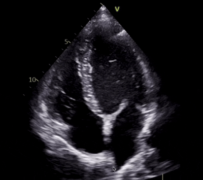
A cardiac ultrasound test that checks the structure and function of the heart muscle, valves, and chambers. A hand-held wand is placed on the chest and high-frequency sound waves are used to create 2D diagnostic images. Doppler and Color Doppler are also used to evaluate blood flow across the heart valves.
-
The out-of-pocket cost for an echocardiogram depends on your insurance plan and deductible.
Across Maine, prices can range anywhere from $800 to $4,300, depending on where the test is performed.
Because hospital-based imaging often includes multiple billing layers, many patients are turning to independent centers for more transparent and affordable pricing.(At Echo Imaging Solutions, our self-pay echocardiogram is a flat $450, with no hidden fees or surprise bills.)
-
There is generally no preparation needed for an echocardiogram. Patients can eat, drink, and take their medications before the test.
You will be asked to remove your clothing from the waist up and put on a hospital gown. Three electrodes will be placed on the chest and you will be lying on your left side for a majority of the echo. A small amount of gel will be put at the end of the transducer and the probe will be placed on different areas of the chest to acquire images.
The sonographer may have you hold your breath to improve the quality of the ultrasound images. The only discomfort may be from the pressure of the probe on your chest.
-
A transthoracic echocardiogram (TTE) can take 40-60 minutes. The length of the echo test can vary depending on the quality of images and degree of findings. We perform this test at our office in South Portland, Maine.
-
Many patients refer to an echocardiogram as an "echo" and this test is used to visualize the heart in real-time. Moving 2D images of the heart are taken to assess the overall structure and function. This is a non-invasive test that is readily available and affordable.
An electrocardiogram (EKG) is used to assess the heart's electrical activity and is not a form of imaging. An EKG produces a graph that displays the heart rate and rhythm.
-
Providers often order a transthoracic echocardiogram to diagnose, prevent, and monitor cardiovascular disease. This test is also common for pre-operative clearance.
Some common reasons for routine echocardiograms are hypertension, atrial fibrillation, heart murmur, aortic stenosis, mitral regurgitation, heart failure, cardiomyopathy, or previous heart attacks.
Visit Us
144 Thadeus Street Suite #1
Portland, ME 04106
Hours
Monday–Friday
8am–5pm
Fax
(833) 972-4822